Facilities
In vision science we make use of the following well equipped facilities:

Vision and Mobility Laboratory
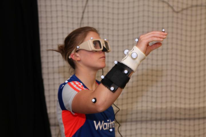
The Vision & Mobility lab, funded by Department of Health, The Health Foundation, PPP Healthcare and NIHR, is a state-of-the-art research facility which allows the evaluation of visual contributions to gait and mobility, including stair negotiation.
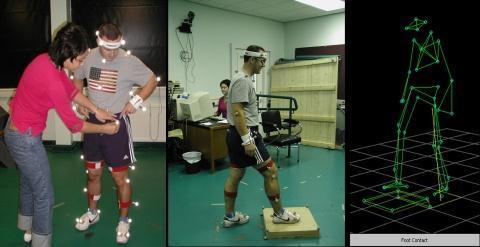
Subjects are fitted with reflective motion tracking tags, then step onto a small platform and the motion-tracker uses the information to reconstruct the subject’s position and stance (see image).
The lab quantifies human movement using a 6 camera Vicon MX3 motion analysis system, and 2 AMTI force platforms. Adaptations in movement pattern are quantified using various biomechanical models.

Adaptive optics laboratory
The Adaptive Optics (AO) laboratory, funded by EPSRC, houses the world’s first binocular adaptive optics system for the study of accommodation and oculomotor function. Funding from the BBSRC supports a
In the AO Lab, optometrists and physicists work together using a unique binocular wave-front sensor to analyse the higher order aberrations of the eye and their consequence for visual function. This group also uses adaptive optics to image the in vivo human retina.
Adaptive optics
Our adaptive optics system uses a deformable mirror to correct higher-order aberrations. The system can be used to effectively by-pass the imperfections in the optics of the eye, thus improving visual acuity. We are currently using this system to investigate the potential role of ocular aberrations in accommodation accuracy during nearwork.
Binocular wavefront sensing
The performance of the human eye is limited by a number of optical and neural factors. Our research laboratory is interested in the measurement of higher-order ocular aberrations (i.e. those above sphere and cylinder). These higher-order ocular aberrations are known to fluctuate over time, and they may be involved in accommodation control.
Our laboratory has facility for the measurement of higher-order aberrations, and their fluctuations, in real time. We are interested in differences in aberration dynamics that may exist between emmetropes and myopes, and stable versus progressing myopes.
Transcranial Magnetic Stimulation (TMS) laboratory
Funding from the BBSRC supports a Transcranial Magnetic Stimulation laboratory, a facility for the fMRI-guided neuro-stimulation of the human brain. This allows the combination of neuroimaging (fMRI), neurostimulation (TMS) and psychophysical methodologies in the examination of human brain function.
We now have the ability to use functional and structural MRI data to accurately guide the delivery of TMS to specifically identified regions of the human brain. Using standard activation paradigms we can identify regions of the brain that are activated by moving stimuli. In addition, we can also use retinotopic mapping procedures to map boundaries of visual areas that are found in the brain. The combination of fMRI data with high resolution structural scans has enabled us to confidently localise and identify, in individual brains, different cortical areas (e.g. V5/MT, V3A and V3B) that have been implicated in the cortical processing of moving visual stimuli. This is important because of the high degree of inter-subject variability in the position of these different areas. Based upon these data we can then position the TMS coil at precise regions of the scalp using a frameless stereotactic co-registration system3. This enables us to accurately target the delivery of TMS to specific visual areas, such as V5/MT+ for example, whilst observers perform behavioural tasks designed to investigate different aspects of motion perception.
Visual electrodiagnostic testing unit
The Visual Electrodiagnostic Testing Service (VETS) is located within the Bradford School of Optometry & Vision Sciences (BSOVS) at the University of Bradford. Our aim is to provide a high quality visual electrodiagnostic testing service for ophthalmologists in the W. Yorkshire region which is embedded into the existing academic research and clinical infrastructure of the University.
The service provides the full range of diagnostic tests that are typically required by ophthalmologists in their diagnosis and management of people with a variety of hereditary and acquired disorders of the visual system. Tests performed include: visual evoked potential (VEP) recording, pattern and flash electro-retinography (PERG and ERG) and electro-oculography (EOG). All testing protocols conform to current clinical (ISCEV) standards.
Vision and Reading Clinic
The Vision and Reading Clinic specialises in the detailed visual assessment for children and adults who are having difficulties with reading.
Difficulties may result from uncorrected refractive error, poor co-ordination and/or focusing of the two eyes, or difficulties when viewing pages of black text printed upon white paper (often termed Pattern Glare, Visual Stress or Meares-Irlen syndrome). Whilst these problems can be found in children and adults with specific learning difficulties such as dyslexia, they can also affect individuals without specific learning difficulties.
Visual symptoms associated with difficulties reading may include some or all of the following:
- Blurred text
- Words appearing to move
- Text appearing to shimmer
- Words fading then reappearing
- Illusions of colour may be seen within the text
- Individuals may also experience sore eyes or headaches
- Individuals may have difficulty following sentences and may lose their place whilst reading
What to expect
Binocular vision assessment
When a patient comes for an assessment in the Vision and Reading Clinic they will have a full binocular vision assessment to look for any anomalies or dysfunction of the focusing and movement systems of the eyes. This will take between 1-2 hours. If any binocular vision anomalies are found they can be managed with eye exercises and/or a spectacle (or contact lens) correction. Follow-up appointments may be needed over a period of a few weeks or months.
Coloured Overlays and Colorimetry
After binocular vision has been assessed and any required treatment prescribed, a coloured overlay assessment may be recommended if the patient is still symptomatic. Coloured overlays are A4 sheets of acetate plastic in ten different colours which can be combined to produce further colours. The overlays are designed to be placed flat on top of reading material to aid reading and they may help alleviate some or all of the symptoms detailed above in some individuals.
If overlays are found to be beneficial and are used successfully by an individual for a period of 3 months or more, a colorimetry assessment is recommended, to ascertain the specific lens colour required for spectacles to give the same effect as the overlay. The use of tinted spectacles is often found to be more convenient when viewing reading material from various sources such as VDU screens and for writing.
Important Point
Whilst we can help alleviate some or all of the visual symptoms experienced by many patients with dyslexia, we are not able to assess or treat dyslexia or any other Specific Learning Difficulty. We do see many individuals with these problems and the aim of the Vision and Reading Clinic is to minimize the effects of any visual problems associated with reading. We cannot cure dyslexia but we can help a proportion of individuals overcome some or all of the visual problems that further hinder their reading capability.
